Diagram Of The Muscles In The Forearm / Human Muscles Diagram : human muscle system | Functions ... / 12 (4 superficial + 3 mobile wad + 5 deep).
Diagram Of The Muscles In The Forearm / Human Muscles Diagram : human muscle system | Functions ... / 12 (4 superficial + 3 mobile wad + 5 deep).. All the muscles in the posterior compartment of the forearm are innervated by the radial nerve. It is a functionally important muscle that contains two heads. Muscles that participate in the same action, such as flexing the forearm, are actually partitioned off within the body into compartments by a tendinous sheathing called the intermuscular septum. The forearm is a mass of some 20 different muscles. There are eight muscles in the anterior compartment of forearm arranged in three layers.
The forearm is a mass of some 20 different muscles. Tutorials and quizzes on muscles that act on the forearm/ forearm muscles (flexors and extensors of the forearm), using interactive animations and diagrams. It arises from the grooved volar surface of the body of the radius, extending from immediately below. 4, attachment… the muscles of the back forearm. Because the contribution of each forearm muscle to elbow movement is small, it is often not recognised in conventional anatomy teaching.
The superficial extensors of the forearm are the brachioradialis, extensor carpi radialis longus, anconeus, extensor carpi radialis brevis, extensor carpi ulnaris, extensor digitorum and extensor digiti minimi.
The forearm is the region of the upper limb between the elbow and the wrist. Remembering the action of each one can be quite difficult. Longus, brevis, longus, brevis (longus is lateral to brevis). Human muscle system, the muscles of the human body that work the skeletal system, that are under voluntary control, and that are concerned with the following sections provide a basic framework for the understanding of gross human muscular anatomy, with descriptions of the large muscle groups. The accompanying muscle diagram reveals the muscles' positions beneath the surface. The antibrachial or forearm muscles may be divided into a volar and a dorsal group. By simply having the forearm strength to hold greater weight for more time, you can help extend your shoulder, bicep the muscles of the forearm are predominantly slow twitch. The forearm is the region of the upper limb between the elbow and the wrist. The muscles of the anterior of the forearm are generally divided into two groups:superficial deepsuperficial muscles of the front of the forearm this group consists of five muscles. The flexor pollicis longus is situated on the radial side of the forearm, lying in the same plane as the preceding. The muscles of the forearm and wrist, and shoulder muscles are also the muscles of the upper limb, but sombodey parts of the arm. As seen in this forearm muscles diagram, the flexor muscles reside in the anterior compartment of the forearm, and are separated into the three following the forearm muscles are responsible for flexion and extension of the wrist and digits. Another handy relation to keep in the back of head is:
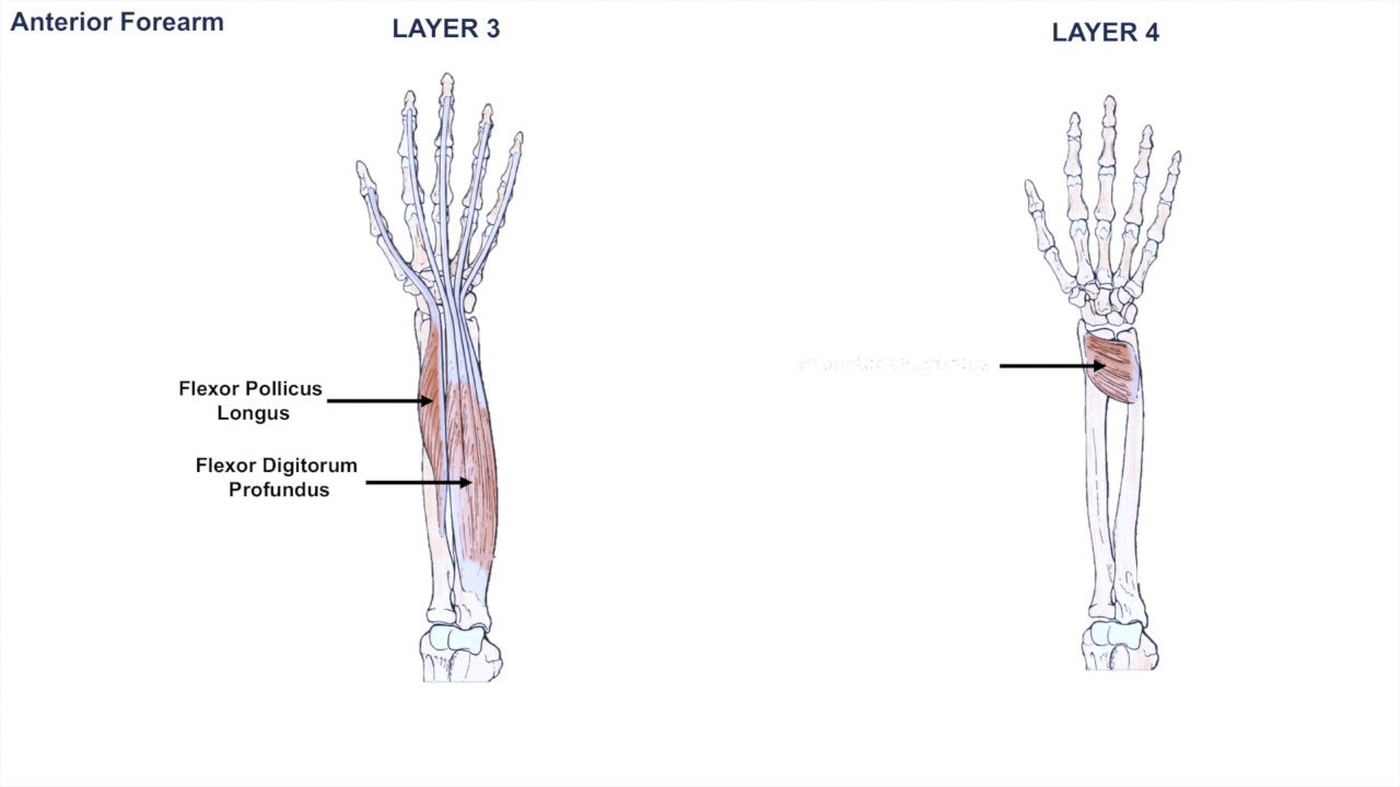
There are eight muscles in the anterior compartment of forearm arranged in three layers. Pronator teres pronates the forearm, turning the hand posteriorly. 12 (4 superficial + 3 mobile wad + 5 deep). This layer contains only one muscle, the flexor digitorum. Inflammation of this region caused by repetitive.

This is the most medial of the superficial flexor muscles in the forearm.
The forearm is divided into two compartments, which are separated by the radius and ulna and the interosseous membrane running between them. Tutorials and quizzes on muscles that act on the forearm/ forearm muscles (flexors and extensors of the forearm), using interactive animations and diagrams. So, the muscles of the anterior compartment are generally innervated by the median nerve, with a few muscles being innervated by the ulnar nerve. The forearm is the region of the upper limb between the elbow and the wrist. Diagram of the muscles of the arm in action. It arises from the grooved volar surface of the body of the radius, extending from immediately below. Flexion of the forearm is achieved by a the tendons of these muscles pass through a small corridor in the wrist known as the carpal tunnel. Inflammation of this region caused by repetitive. Remembering the action of each one can be quite difficult. Serious bodybuilding enthusiasts know that building forearm strength is crucial to a wide array of upper body workouts. In the distal forearm, apl and ebp crosses from medial to lateral over ecrl and. Superficial muscles of the posterior forearm: This layer contains only one muscle, the flexor digitorum.
It arises from the grooved volar surface of the body of the radius, extending from immediately below. Remembering the action of each one can be quite difficult. Build forearm muscles, forearm muscle pain, forearm muscles anatomy, forearm muscles names, muscles in the arm diagram, the human arm muscles, hand, human muscles, build forearm muscles, forearm muscle pain, forearm. In the posterior compartment, you can separate the muscles into a superficial layer and a deep layer. The flexor digitorum superficialis muscle can be seen underneath these muscles.
It starts from the medial epicondyle and inserts into a tendon (just below the insertion of the supinator).
Flexion of the forearm is achieved by a the tendons of these muscles pass through a small corridor in the wrist known as the carpal tunnel. There are many muscles in the forearm. Diagram the movements of the humerus muscles that act on the forearm. It is a functionally important muscle that contains two heads. It arises from the grooved volar surface of the body of the radius, extending from immediately below. The flexor pollicis longus is situated on the radial side of the forearm, lying in the same plane as the preceding. The forearm is the region of the upper limb between the elbow and the wrist. The antibrachial or forearm muscles may be divided into a volar and a dorsal group. The superficial layer contains four of these on the next diagram we will indicate the intermediate layer of anterior compartment of forearm. 12 (4 superficial + 3 mobile wad + 5 deep). In the anterior compartment, they are split into three categories: The accompanying muscle diagram reveals the muscles' positions beneath the surface. Another handy relation to keep in the back of head is:


Komentar
Posting Komentar